
.
Replication fork
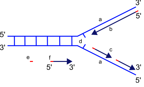
Scheme of the replication fork. (*)
a: template, b: leading strand, c: lagging strand, d: replication fork, e: primer, f: Okazaki fragments
The replication fork is a structure that forms within the nucleus during DNA replication. It is created by helicases, which break the hydrogen bonds holding the two DNA strands together. The resulting structure has two branching "prongs", each one made up of a single strand of DNA. These two strands serve as the template for the leading and lagging strands which will be created as DNA polymerase matches complementary nucleotides to the templates; The templates may be properly referred to as the leading strand template and the lagging strand template.
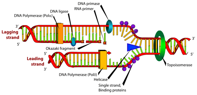
Many enzymes are involved in the DNA replication fork.
Replication
When replicating, the original DNA splits in two, forming two "prongs" which resemble a fork (hence the name "replication fork"). DNA has a ladder-like structure; imagine a ladder broken in half vertically, along the steps. Each half of the ladder now requires a new half to match it. Because DNA polymerase can only synthesize a new DNA strand in a 5' to 3' manner, the process of replication goes differently for the two strands comprising the DNA double helix.
Leading strand
The leading strand template is the template strand of the DNA double helix that is oriented in a 3' to 5' manner. All DNA synthesis occurs 5'-3'. The original DNA strand must be read 3'-5' to produce a 5'-3' nascent strand.
The leading strand is formed along the leading strand template as a polymerase "reads" the template DNA and continuously adds nucleotides to the 3' end of the elongating strand. This polymerase is DNA polymerase III (DNA Pol III) in prokaryotes and presumably Pol ε[1][2] in eukaryotes.
Lagging strand
The lagging strand template is the coding strand of the DNA double helix that is oriented in a 5' to 3' manner. The newly made lagging strand still is synthesized 5'-3'. However, since the DNA is oriented in a manner that does not allow continual synthesis, only small sections can be read at a time. An RNA primer is placed on the DNA strand 3' to the origin of replication. Just as before, DNA Polymerase reads 3'-5' on the original DNA to produce a 5'-3' nascent strand. Polymerase reaches the origin of replication and stops replication until a new RNA primer is placed 3' to the last RNA primer. These fragments of DNA produced on the lagging strand are called Okazaki fragments. The orientation of the original DNA on the lagging strand prevents continual synthesis. As a result, replication of the lagging strand is more complicated than of the leading strand.
On the lagging strand template, primase "reads" the DNA and adds RNA to it in short, separated segments. In eukaryotes, primase is intrinsic to Pol α.[3] DNA polymerase III or Pol δ lengthens the primed segments, forming Okazaki fragments. Primer removal in eukaryotes is also performed by Pol δ.[4] In prokaryotes, DNA polymerase I "reads" the fragments, removes the RNA using its flap endonuclease domain, and replaces the RNA nucleotides with DNA nucleotides (this is necessary because RNA and DNA use slightly different kinds of nucleotides). DNA ligase joins the fragments together.
See also
* DNA replication#The replication fork for context
References
1. ^ Pursell, Z.F. et al. (2007). "Yeast DNA Polymerase ε Participates in Leading-Strand DNA Replication". Science 317 (5834): 127. doi:10.1126/science.1144067. PMID 17615360.
2. ^ Scott D McCulloch; Thomas A Kunkel (01/2008). "The fidelity of DNA synthesis by eukaryotic replicative and translesion synthesis polymerases". Cell Research 18 (1): 148. doi:10.1038/cr.2008.4. PMID 18166979.
3. ^ Elizabeth R. Barry; Stephen D. Bell (12/2006). "DNA Replication in the Archaea". Microbiology and Molecular Biology Reviews 70 (4): 876–887. doi:10.1128/MMBR.00029-06. PMID 17158702.
4. ^ Distinguishing the pathways of primer removal during Eukaryotic Okazaki fragment maturation Contributor Author Rossi, Marie Louise. Date Accessioned: 2009-02-23T17:05:09Z. Date Available: 2009-02-23T17:05:09Z. Date Issued: 2009-02-23T17:05:09Z. Identifier Uri: http://hdl.handle.net/1802/6537. Description: Dr. Robert A. Bambara, Faculty Advisor. Thesis (PhD) - School of Medicine and Dentistry, University of Rochester. UR only until January 2010. UR only until January 2010.
External links
* DNA Replication and Repair

Retrieved from "http://en.wikipedia.org/"
All text is available under the terms of the GNU Free Documentation License
